
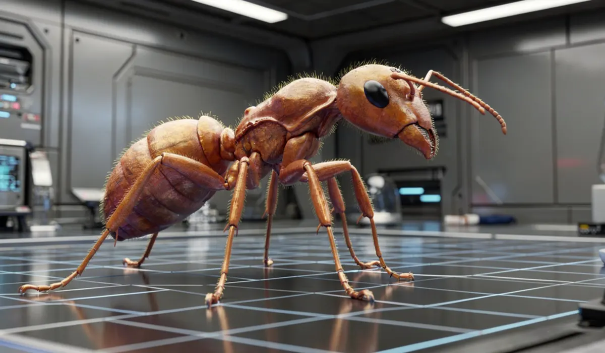
If you think ants are just tiny insects crawling around your backyard, think again. What if I told you that scientists are now capturing their entire bodies in stunning 3D detail, creating digital models so precise you can explore their anatomy from anywhere in the world? This is not science fiction—it’s happening today.
- How High-Throughput Scanning Brings Ants to Life
- The Role of AI in Creating Usable 3D Models
- Building a Global Database of Ant Morphology
- Why AI and Imaging Matter Beyond Ants
- Exploring Ant Anatomy Like Never Before
- Challenges and the Road Ahead
- The Future of Morphological Big Data
- Unlocking the Hidden World of Ants with AI Ant Scan Technology
The process behind this marvel involves more than just fancy cameras. It’s a blend of advanced imaging technology and smart algorithms that turn raw data into rich, interactive 3D models. And yes, artificial intelligence plays a crucial role in making this possible, transforming how we study the natural world at a microscopic level.
How High-Throughput Scanning Brings Ants to Life
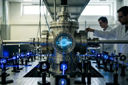
Capturing the intricate structure of ants in three dimensions isn’t as simple as snapping a photo. Scientists use a technique called synchrotron X-ray microtomography, which is basically a super-powered CT scan designed for tiny creatures. This method shoots X-rays through ethanol-preserved ants, generating thousands of images from different angles in mere seconds.
The magic lies in how these images are processed. Thanks to the high flux density of synchrotron radiation, scans reveal not only the hard exoskeleton but also soft tissues like muscles and organs without needing invasive preparation. This level of detail is crucial for understanding ant biology, behavior, and evolution.
The challenge? Millions of ant species exist, and scanning each one manually would take years. That’s where automation comes in. Using robotic sample exchangers, researchers can scan dozens of ants per hour, producing a massive dataset that covers hundreds of genera and species worldwide.
The Role of AI in Creating Usable 3D Models
Raw scan data is complex and bulky. Imagine thousands of 2D X-ray slices stacked like pages in a book, each needing interpretation. AI steps in to make sense of this data. Neural networks trained on annotated scans learn to automatically segment ants from the background, identify body parts, and even separate muscles from exoskeletons.
This automation drastically reduces the time and manual effort required to create 3D models. Instead of experts spending hours on each specimen, AI can process large batches, cropping images and generating surface meshes that are ready for analysis or visualization.
The standardization of scanning parameters is key here. Because all ants are scanned under similar conditions, AI models can be trained more effectively, ensuring consistent results across thousands of specimens. This comparability is essential for large-scale studies on morphology and evolution.
Expert Tip
Standardized imaging conditions enhance AI segmentation accuracy, enabling faster and more reliable 3D reconstructions across diverse ant species.
Building a Global Database of Ant Morphology
The Antscan initiative has created an open-access database with over 2,000 high-resolution 3D scans covering nearly 800 ant species from all over the world. This resource is more than a collection of images; it’s a digital library that links morphology with ecological and genetic data.
Researchers can explore 3D models online, download raw tomograms, or use advanced tools to segment and analyze anatomical features. The database also connects with genome sequencing projects, allowing scientists to correlate physical traits with genetic information—an approach that could unlock new insights into how ants have diversified and adapted.
Making these data freely available encourages collaboration and innovation. It also democratizes access to specimens that would otherwise require physical travel to museums or labs, opening doors for educators, students, and citizen scientists.
Why AI and Imaging Matter Beyond Ants
While ants are the focus here, the technology and methods developed have broader implications. Many small organisms, especially insects, have been difficult to study in 3D due to their size and complexity. High-throughput synchrotron micro-CT combined with AI-driven data processing offers a scalable way to digitize biodiversity.
This approach supports ecological studies, evolutionary biology, and even biomimetic design, where engineers look to nature for inspiration. By capturing internal and external anatomy in detail, researchers can model biomechanics, physiology, and developmental processes with unprecedented precision.
The challenge remains access to synchrotron facilities and the computational resources needed for data handling. However, advances in robotics, imaging optics, and cloud computing promise to expand availability and reduce bottlenecks.
Exploring Ant Anatomy Like Never Before
One striking example from Antscan is the ability to segment individual tissues within ants. For instance, scientists can isolate muscles, nervous systems, digestive tracts, and even the sting apparatus in 3D. These models can be animated or 3D printed, providing tangible insights into form and function.
This level of detail helps answer questions about how ants perform complex tasks, how their bodies have evolved in response to ecological pressures, and how different castes within a colony specialize anatomically. The combination of imaging and AI thus turns static specimens into dynamic subjects of study.
Challenges and the Road Ahead
Despite the progress, several hurdles remain. Specimen preservation varies, which can affect scan quality. Some ants show tissue shrinkage or damage from prior handling, complicating interpretation. Also, scanning larger specimens requires different setups or multiple scans stitched together.
AI segmentation, while powerful, is still evolving. Complex anatomical features may require manual correction or retraining of neural networks. Data storage and transfer are significant concerns given the terabytes of information generated.
Nevertheless, the collaboration across institutions and the open-access model provide a strong foundation. Continued development in imaging technology and AI promises to refine workflows, improve data quality, and extend coverage to more species.
The Future of Morphological Big Data
The Antscan project illustrates how combining sophisticated imaging with AI can scale up the study of organismal form. As databases grow, researchers can conduct comparative analyses across thousands of species, linking morphology with genetics and ecology.
This integrated approach will deepen our understanding of biodiversity, evolutionary processes, and functional biology. It also highlights the importance of open data and collaborative infrastructure in modern science.
For anyone curious about the tiny architects of ecosystems, these 3D models offer a new lens to explore their complexity. And with AI turning raw scans into detailed models, the micro-world of ants is now more accessible than ever.
Unlocking the Hidden World of Ants with AI Ant Scan Technology
The combination of high-throughput synchrotron microtomography and AI-driven data processing has opened a new chapter in biological research. By capturing and converting ant scans into detailed 3D models, scientists have created a resource that goes far beyond traditional specimen collections.
This approach allows for rapid, standardized, and non-destructive digitization of small organisms, preserving their morphology in exquisite detail. It also bridges the gap between physical specimens and digital analysis, empowering a broader community of researchers and enthusiasts.
With the Antscan database and its AI-enhanced workflows, the study of ant biodiversity and evolution is entering a new era. This technology not only reveals the hidden complexity of ants but also sets a precedent for future efforts to document life’s diversity in three dimensions.
How does synchrotron X-ray microtomography differ from regular CT scans?
Synchrotron micro-CT uses a highly intense and focused X-ray beam produced by a synchrotron facility, allowing much faster scans at higher resolution and better contrast, especially for small and delicate specimens like ants.
Why is AI necessary for processing ant scans?
The raw data from scans consist of thousands of images that need to be segmented and interpreted. AI automates this process, reducing manual labor and increasing consistency across large datasets.
Can anyone access the 3D ant models?
Yes, the Antscan database is publicly available online, providing free access to thousands of 3D scans and associated metadata for research, education, or personal interest.
What are some practical applications of these 3D models?
They can be used for evolutionary studies, biomechanical modeling, education, virtual dissections, and even 3D printing for hands-on learning.
Are there limitations to the current technology?
Access to synchrotron facilities is limited, and some specimens may not scan perfectly due to preservation quality. AI segmentation may also require refinement for complex anatomical features.


